Throat model - Pulse MDM

When Models Marry Cadavers

How Hands-On Models and Cadaver Labs Create the Best Experience for Training Medical Professionals

Cadavers have been used for medical training and research since the 13th century. Over the past several hundred years, cadavers have undoubtedly enhanced the medical profession and allowed a mode of training that garnered a realistic and generally reproducible training experience. As technology has progressed exponentially over the years, cadaver labs have been joined by hands-on anatomical models to enhance the training of clinicians, residents, and medical students. To this day, cadaver labs continue to be an extremely effective mode of training but are now joined on the throne of gold-standard medical training by hands-on models.

Cadaver Labs: The Pros

Cadaver labs have stood the test of time. Just ask any patient-facing medical provider or medical student if they’ve worked with a cadaver at least once, and nearly all will tell you that they have.

Although rather eerie, let’s face it, cadaver labs are cool. They provide us with a true-to-scale way to learn about the human body. Along with that, they provide the ability for healthcare providers to train and learn their craft without the risk of negative outcomes.

With the obvious fact that cadavers lack the blood flow of a living patient, they are all tested for pathogens (including Covid) and must meet certifications that the required tissues are present before they can be utilized by their intended clientele.

Cadaver Labs: The Drawbacks

Just as the positive aspects of cadaver labs should be recognized, it is equally important to weigh the shortcomings of this long-standing method of training. First is the availability concern. Unfortunately, cadaver availability for medical and scientific purposes is extremely limited when compared to the need. And for those that do become available, there’s only ever the opportunity for one ‘first cut’ per body structure. This often leaves a room full of clinicians or students standing around watching as one or a few people practice a skill that will soon become unavailable for anyone else to practice.

Even though nothing can be more real than actual human tissue, cadavers are also typically left with features that aren’t particularly realistic. This may sound a bit morbid, but consider that after a person dies, their tissues, organs, and systems no longer have the amazingly orchestrated processes occurring that allow humans to be human. For anyone who has ever been in a cadaver lab, this explains the impossible-to-ignore aroma of embalming chemicals.

Although chemicals can preserve the cadaver from decaying externally, these same chemicals affect the external tissue properties of the cadaver. Additionally, it is impossible to prevent the natural events that occur internally after death. Even with a short turnaround time, many of the internal and external tissue properties of the cadaver will no longer resemble that of the living patients clinicians will work with.


The New Age

With the incredible technology we have available to us today, medical device manufacturers, medical salespeople, and clinical trainers can help fill in the cracks that cadaver labs leave behind. Nearly all the shortcomings of cadaver labs outlined above can be supplemented by hands-on anatomical models.

Today, hands-on models are being adopted as either an alternative or supplement to cadavers by some organizations but continue to be overlooked by others. The reasons for those that overlook the models vary but generally comes down to either a lack of awareness of the versatility of hands-on models or a belief that models do not provide a high-quality, realistic experience. In reality, models are fully customizable for the client, ensuring they are built to the specific standards necessary to provide the exact training experience needed for their healthcare providers.

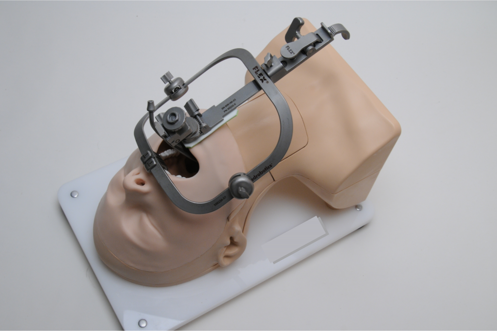
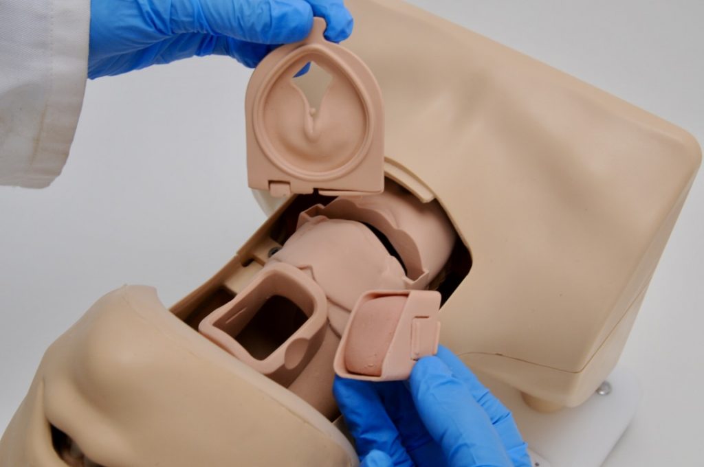
As outlined in last month’s blog post, hands-on models are designed and cast with extreme dedication to replicate specific clinical scenarios. The design, materials, colors, and transparency can all be altered until they meet the exact specifications of the client. This eliminates the issue found with cadaver labs where the cadaver’s structures no longer resemble a realistic feel or durability of a living patient.

More importantly, the “one first cut” problem is completely eliminated with cleverly designed consumables. As discussed above, the limited availability of cadavers often leaves many students and clinicians in a position where they are doing more observing of their peers than actually performing incisions and surgical techniques themselves. Custom-designed hands-on models provide easily replaceable parts in any quantity needed to ensure that every surgeon can make that first cut to receive the proper training and experience needed for proficiency and then utilize cadavers if necessary to perfect the technique on human tissue. This is a telling example of how cadaver labs and hands-on models can supplement one another to provide the best training experience for clinicians and students.

Cost Comparison

From the perspective of a healthcare organization, the level of reusability and transportability that a model provides means significant cost savings over the long term by eliminating nearly all the recurring costs necessary for cadaver labs. That’s, of course, not to say that both options don’t have their associated costs. A general breakdown of the costs of each looks like this:


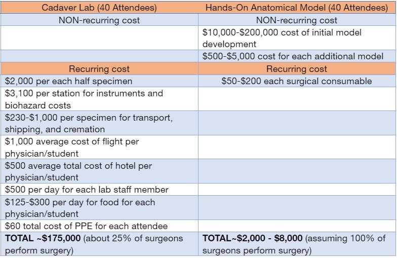
Although both cadaver labs and hands-on models require initial costs, the savings from the lack of recurring costs of hands-on models make the “break-even” point quickly realized.

Cadavers and hands-on models both clearly have their place in medical/surgical training. With healthcare organizations aiming to remain as profitable as possible while simultaneously ensuring their providers can access the highest level of training for improved patient outcomes, this is where the skill and knowledge of medical device manufacturers, medical device salespeople, and clinical trainers truly comes into play. Based on the outlined benefits of hands-on models, it is easy to see how discussing this option with healthcare organizations creates some obvious selling points. In addition to what has been outlined above, note the following:


Hands-on anatomical models provide the following benefits and selling points for your clients:

  • Able to be mono or bipolar ablated, lasered, stapled, injected, dissected, debrided, and cut
  • Infinite custom pathologies that cadavers never have
  • Unlimited anatomical variation
  • Nearly zero recurring costs once the development and unit costs are covered
  • No team required to set up off-site cadaver training
  • No team required to make travel arrangements for clinicians
  • No clinician travel, lodging, meal, or entertainment costs
  • No lab rental costs
  • No lab technician costs
  • No cadaver shipping costs
  • No surgical instrument sterilization
  • No cadaver disposal costs
  • Training the entire clinical staff as opposed to just the main surgeon by meeting your clients at their locations with a portable model with replaceable parts.
  • Consistent and repeatable experiences. Every surgeon gets to make the 1st cut using low-cost, easily replaceable surgical inserts.
  • Increased training opportunities since the model can be used and set up without a lab and can be done anywhere (like a hotel conference room).

More training sessions = more mastery.
More mastery = more confidence in your device/procedure.
More confidence = increased surgeon adoption of your procedure.


Wrap-Up

Allowing every surgeon to make the first cut and to hone their skills with a wide range of pathologies and anatomical variance is essential for the safety of the patients whose lives are in their hands every day. By using models as precursors to cadaver labs or in tandem with cadavers, medical students, residents, and clinicians new to a particular type of procedure gain the repetition and muscle memory necessary to perfect their skills, so they have the confidence and buy-in of a technique much more quickly. These models can then be used within patient appointments to ensure the patient has a clear understanding of what is involved in their specific procedure. Getting clinicians to this point of confidence and mastery requires an important role from medical device manufacturers and clinical trainers. The success of your mission comes from the success the surgeons have with their patients.


Pulse Medical Demonstration Models – Making Dreams Come True

When you’re in the model-making business, it’s a true joy to help our clients find the perfect solutions to their greatest challenges. Because our models are customized exactly for your need, we are a huge game-changer for those who need quality models. Contact us today to see how we can help you!

3D Machine - Pulse MDM

Should I Use 3D Printing For My Anatomy Models?

Should I Use 3D Printing For My Anatomy Demonstration and Training Models?

Although 3D printing originated in 1984, the technology has caught fire in the past 15 years or so. It’s become a buzzword in many industries involved in engineering and design (and, more recently, medicine). With all of the publicity surrounding it, it’s reasonable to wonder, “is 3D printing the best option for my anatomy models?” For that answer, let’s dive a little deeper.

3D Printing: How It’s Used

3D printing is used for anatomy models! For hard structures, such as bone models, 3D printing may be the sole process used to create the finished product. For tissues of the body, however, there are varying levels of elasticity and softness. After all, most structures of the human body are comprised of soft tissue, not rigid like bone. This is where things get a little more interesting.

To create 3D-printed anatomy models, there has to be an “outline” to define the shape and size. For this, 3D CAD (computer-aided design) data is created of the anatomical structures. This CAD data can be created by using patient data as a starting point or a combination of digital sculpting and engineering software.

After the CAD outline is finalized and accurate to replicate the anatomical shapes and required pathologies, the power of 3D printing is utilized. A prototype (known as a “master”) is 3D printed using the CAD data to allow for a physical product to be visualized for the size and dimensions of a model for client review and approval.

If everything looks good, this master is used as a master pattern for a soft mold to be created around it. This mold brings us to where the magic really happens.

3D Printing: How It’s NOT Used

As beautiful as the 3D printed master is, it’s rigid and therefore not representative of soft tissue. In order to produce a clinically relevant, reusable, durable, and economic soft tissue model, soft materials are cast into the soft mold created from the 3D-printed master model.

This process is highly customizable because the recipe for the soft cast materials can be adjusted repeatedly using the same mold until the exact desired soft tissue feel is created. This is not unlike using the same muffin tin one day to make corn muffins, the next day chocolate cupcakes, and mini quiches the day after. The recipe is different, but the shape remains faithful to the original 3D data/master/mold shape.

When the correct mixture of soft materials is finalized to match the desired clinical scenario, you’re left with a visually beautiful and anatomically accurate model in shape and feel.

Bringing It All Together

Think about 3D printing as a tool that leads to a beautiful final product made of soft materials. The 3D-printed master allows for wonderful visualization, as well as the ability to act as the pattern for a soft mold to be created around it. However, this rigid version of the model simply won’t do as the final product if you wish to provide the most clinically relevant experience while remaining economically responsible.

Rigid 3D printed models require significant labor time for sanding, they cannot replicate the elasticity, responsiveness, and feel of human soft tissue, and they simply don’t last as long as soft cast materials before the color and material properties start to fade. There are competing technologies in 3D printing, but they do not meet the intended purposes of a quality medical demonstration model.

For instance, 3D printing with cells or gels is technically an option, but this has even more limitations than rigid models. While 3D cell printing may be advantageous for research and development purposes where a high degree of tissue property realism is key and convenience is not a requirement, it is not ideal for demo and training uses because it is costly, requires a biologist, are difficult to maintain and has a short shelf-life.

3D printing soft materials is another technology. While this option has a longer shelf-life than cell and gel printing (or cadaver parts) and eliminates the issue of only being able to print rigid structures, it significantly lacks clinical relevance because the product does not feel or behave like soft tissue. Additionally, this process leaves a 3D print texture that cannot be sanded out due to its soft nature.

For it all to come together, soft casting materials are the best combination of realism, economy, and convenience.

Soft cast models are better than 3D printed parts in the following ways:

More economical.

  • The finishing work is done once for the master, so there is minimal labor to create more parts.
  • You can make more models in less time.
  • Cast parts are more durable and will last much longer, precluding the need to replace them.

More convenient.

  • These models do not need to be kept in a very specific temperature range like 3D cell printing models and cadaver parts do, making them easier to transport and ship.

More repeatable.

  • They’re created from a mold (and therefore identical), so they’re able to deliver the same experience every time.

More professional.

  • These models have a clean, professional appearance to reflect well on your brand.

With all of your newfound knowledge, let’s circle back to the original question: “Should I use 3D printing for my anatomical models?”

Yes. Just not for all of it.

medical device training - Pulse MDM

What’s Next in Medical Device Marketing & Training

Nobody could have predicted how the pandemic would uproot medical device marketing and training. But there’s one prediction we can safely make today: The hoops we jumped through to keep sales reps engaged and trainers training—from their homes—did more than shift us to virtual meetings and presentations. It kicked open the door to new ideas that medical device makers can use today and after the pandemic.

But those new ideas aren’t necessarily obvious. Clients tell me they’re concerned that this new world order, where they’re limited to conferencing, animations, videos, and other forms of purely digital interaction, suffers from the loss of physicality. They’re investing more in digital but are concerned about reduced results due to the loss of physicality; hands-on, clinically relevant interactions that transfer knowledge viscerally to sales teams and clinicians.

This doesn’t have to be the case. Rather than look at medical device marketing as an either/or proposition—either clinically realistic physicality or 100% digital—we can bring these worlds together with a new hybrid approach. It’s possible to create unparalleled experiences by fusing digital with real-world custom anatomical medical models and set the stage for more effective medical device marketing and training that delivers returns long after the pandemic is over.


BEYOND SHOW AND TELL

The history of disruptive innovation has lessons for us. Look no further than telehealth, which last year erupted from a small number of early adopters to a primary healthcare delivery channel. Those indoctrinated into telehealth won’t abandon it after the pandemic, but they won’t be using it exclusively, either. A hybrid approach is emerging where telehealth consumers are blending virtual check-ins with physical visits.

How can you blend hands-on physical experiences and virtual interactions to deliver a more effective hybrid solution to your sales teams and clinicians? Think beyond show and tell. Think show, tell, and feel. Think about what you would do if you had a clinically responsive realistic physical model to incorporate into your virtual presentations. Instead of people multitasking while you’re presenting, think about your participants interacting with a hands-on model you sent them ahead of time. Imagine the model has been engineered to produce the specific “aha!” moment you need to drive adoption.

Here’s one of many examples I can share based on actual engagements I’ve had with Pulse MDM clients. Sclerotherapy is a procedure used to treat varicose and spider veins.  The doctor injects a solution (normally a salt solution) directly into the diseased vein, causing the vein to close, and forcing blood to reroute through healthier veins.


THE POWER OF TOUCH

Sounds straightforward, but there’s a problem: It takes skill to find and treat tiny varicose veins; a skill that is impossible to acquire using purely digital techniques. The solution we came up with: Adding physicality to the virtual world. We helped our client develop an operable custom model that enhances digital training and sales presentations with a truly tactile experience.

The sclerotherapy model’s vessels contain blue food coloring in the vessels. When clinicians target and inject properly, the blue liquid is chased away by clear water, making the vessel lumen under the skin invisible, giving the appearance of the vessel closing.

Now our client can provide an interactive hybrid experience that uses digital video instruction married with real-world physicality to show, tell, and feel. Clinicians can feel the experience of finely targeting small varicose veins and feel the satisfaction of doing so successfully. This provides a level of enlightenment and confidence no digital technique can match.

This is only one example of how you can create a hybrid “show, tell, and feel” experience for sales and training that delivers better results than show and tell alone. In the end, it’s the difference between giving your clinicians models you point to versus models they touch and experience. Frankly, it’s the only way to ensure you deliver that “aha!” moment in today’s marketing and training environments.


Pulse Medical Demonstration Models Offers Flexible Solutions

It’s not an either/or decision. Here at Pulse Medical Demonstration Models, we have learned it’s a “best of both worlds” decision to blend what we’ve learned about using digital tools during the pandemic with what we know about using physically engaging anatomical medical models. Those who embrace this hybrid approach to medical device marketing and training will gain a distinct competitive advantage over those who fail to evolve. Contact Pulse today to learn more!

Barbie face and body suit - Pulse MDM

It’s More Important to Feel Good than to Look Good

Barbie looks good. But when it comes to training physicians with new medical devices and procedures, how the medical demonstration model feels is more important.

Those hard plastic models we’ve all seen in physicians’ offices look interesting, but you can’t do anything with them. The model pictured above was developed to train clinicians on a non-invasive fat cell freezing procedure and provide hands-on experience of working dimpled, pliable, fibrous, uneven, lax and stiff fat, among others.

We specialize in materials development to model tissue that, short of a cadaver, precisely replicates the response of a human organ to a needle, ultrasound, scalpel, staple, or a myriad of other medical procedures.

Don’t get me wrong, Barbie’s great. And when she gets through medical school, we’ll be right there to help with her training.

I invite you to review our portfolio or to contact me directly to discuss how a medical demonstration model can help you and meet the new challenges of the training environment.

Enthusiastically,
Allison Rae
267-789-6515/ Allison@PulseMDM

Shrimp vs. PulseMDM - Pulse MDM

Shrimp vs. The Medical Demonstration Model

Shrimp

Medical Demonstration Model

√  Barbecue it √  Scope it
√  Boil it √  Ultra-sound it
√  Fry it √  Suture it
√  Saute it √  Bandage it
√  Sear it √  Ablate it
√  Broil it √  Squeeze it
√  Stew it √  Stent it
√  Inject it
√  Pressure it
√  Navigate it
√  Expand it
√  Palpate it

The Pulse MDM Medical Model Wins for Versatility!


Even Bubba Gump knows the days of a hard plastic, painted model sitting on a physician’s desk or in the waiting room are over.  Today, models can be used for advanced clinical training and sales enablement.

What makes medical demonstration models so effective is they offer a tactile experience and are designed to replicate the feel of human organs.  And because of their life-like qualities, medical demonstration models can offer the opportunity to perform procedures on them such as ablation or suturing or navigating through an obstruction that is expected and necessary for any training program.  In many cases, these models can be developed to replace the need for cadaver labs.

Bubba Gump thinks shrimp is pretty versatile, but it’s not as versatile as a Pulse MDM medical demonstration model.

I invite you to review our portfolio or to contact me directly to discuss how a medical demonstration model can help you and meet the new challenges of the training environment.


Enthusiastically,

Allison Rae

267-789-6515/ Allison@PulseMDM

High Tech Vs. High Touch - Pulse MDM

High Tech Vs. High Touch

In his seminal work Future Shock, author Alvin Toffler discoursed on the theme of high tech versus high touch.  Toffler posited that as technology-saturated our lives, we would increasingly seek opportunities for face to face, human interaction.

Continue reading