Built For the Job
Today, we arrive at the fourth and final installment of our series that aims to answer the question, “Do all medical demonstration models need to represent anatomy?” (Spoiler: The answer is no).
In the first three articles of the series, we discussed how and why device replicas are used, how medical skills training can be turned into a game, and whisked you away to a fictional land called Devicia to shed some light on an important training and demo opportunity.
Today, we return from Devicia back to the homeland to wrap the series up by conveying how certain models are built to display the effectiveness of medical devices without the need to imitate the anatomy they are used for in practice.
Devices like the ones we will highlight below help medical device companies demonstrate the amazing attributes of their products as they perform their intended tasks. The goal of these device manufacturers in these instances is to create a medium to train and demonstrate their devices in a way that will optimize skill acquisition and visualization of the intended task without the need to make that medium closely align with the human anatomy it will be performed on clinically.
Get Your Fix
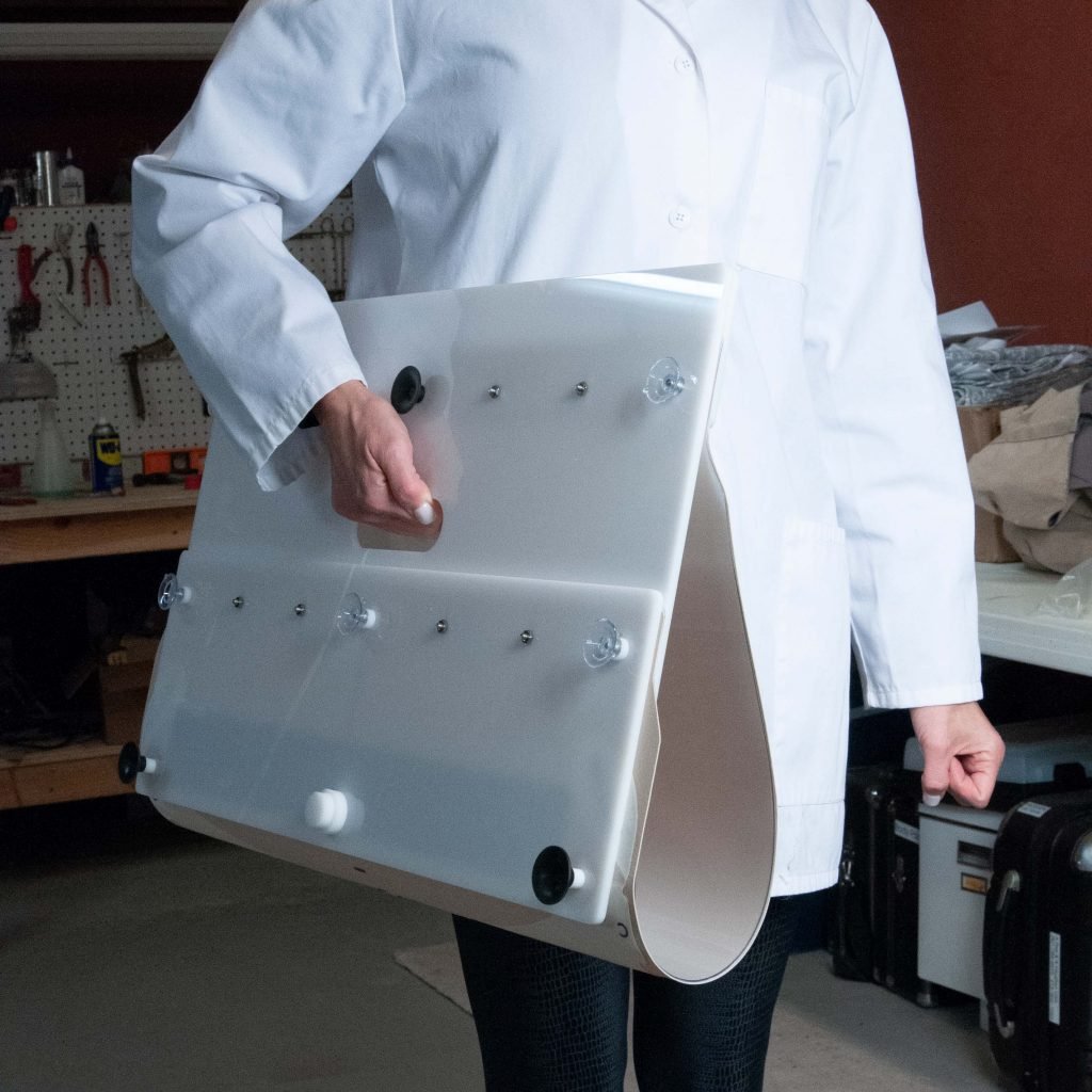
As we have talked about in previous newsletters, portability and convenience is a key part of what we do. From the viewpoint of a medical salesperson or trainer, a product that can’t be demonstrated or trained to its full potential is a product that is more difficult to sell.
As discussed in part one of this series, there are numerous instances where a product needs to be scaled in a way that allows it to be more easily transported or demonstrated than the actual product would allow for. Sometimes this means scaling a very large product down or only including the necessary components for demonstration or training. Other times, this means creating a replica of a product that allows for a more convenient visualization of its functionality.
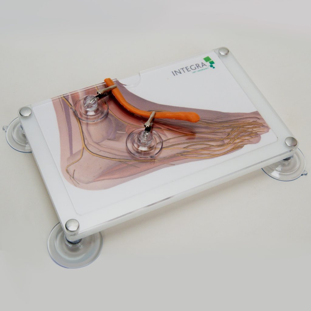
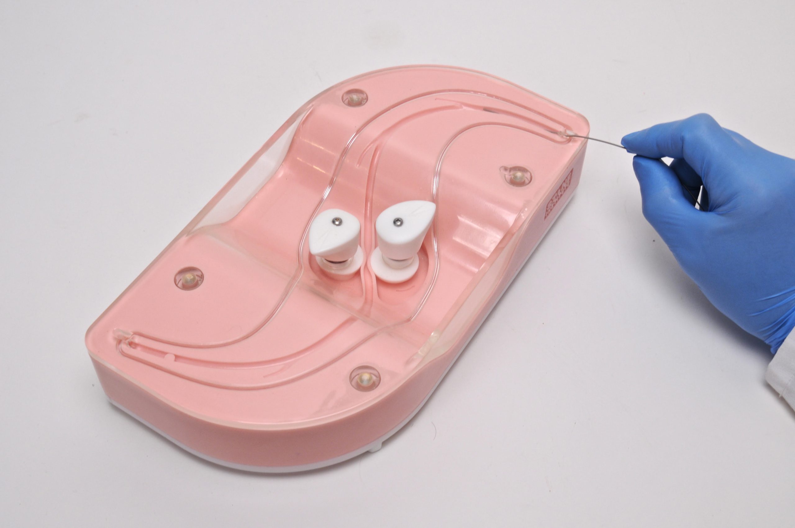
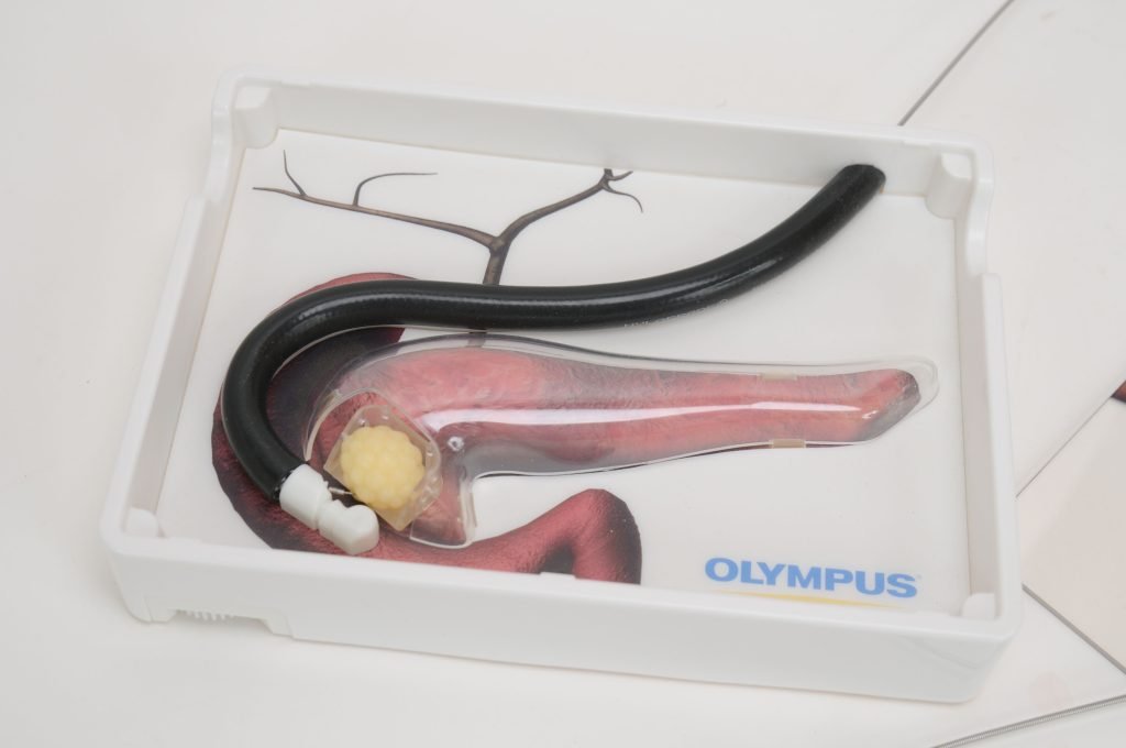
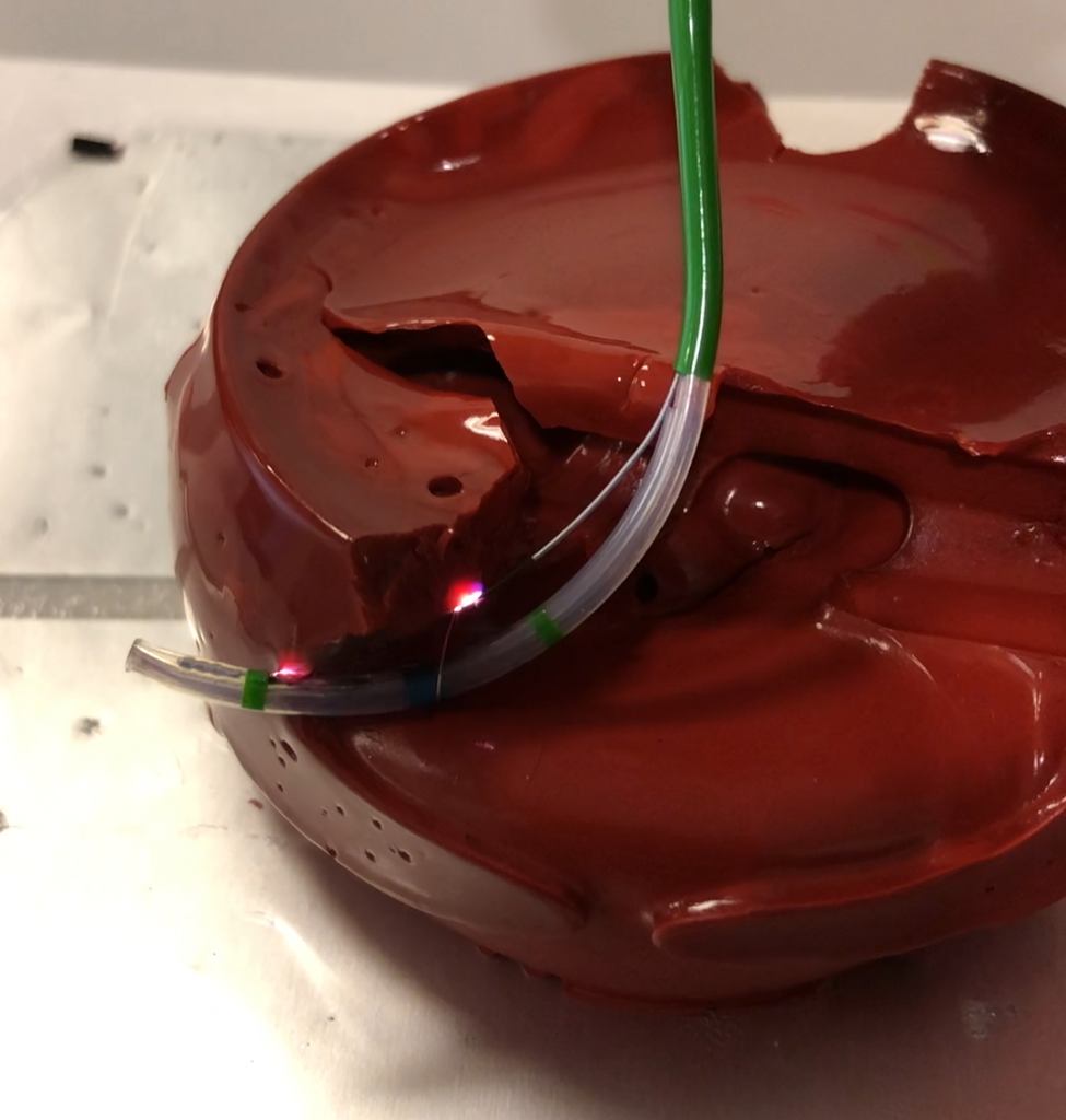
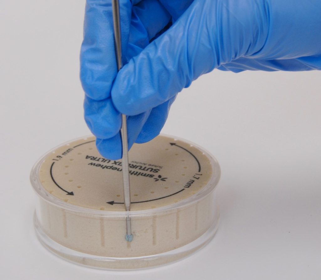
An example that touches on both aspects is a replica we created for a Smith & Nephew product called the Suturefix Ultra. This product is an all-suture fixation device used primarily during rotator cuff repair and hip/shoulder labral repair procedures to provide a durable and secure anchor while removing less bone than traditional fixation anchors. It also utilizes a click-in-place audio cue for the surgeon to confirm the anchor is set.
The challenge our customer was looking to solve was finding a way to demonstrate the advantages of the Suturefix Ultra in a way that would allow both method-of-action visualization of what was occurring in the patient’s joint during the procedure, as well as testing its hold strength and experiencing the tactile feedback.
For this challenge, the best solution was not to create an anatomical model of the shoulder or hip, as this would have resulted in a few shortcomings:
- It would have implied the device could be used for the shoulder and hip only.
- It would have vastly reduced the number of practice sites available in the model.
- It would have reduced visualization.
- It would have decreased portability.
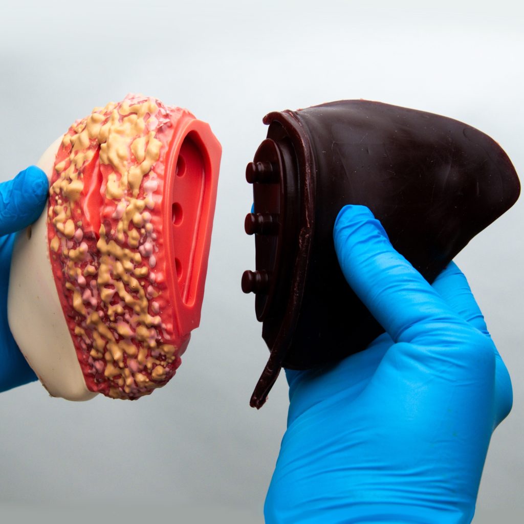
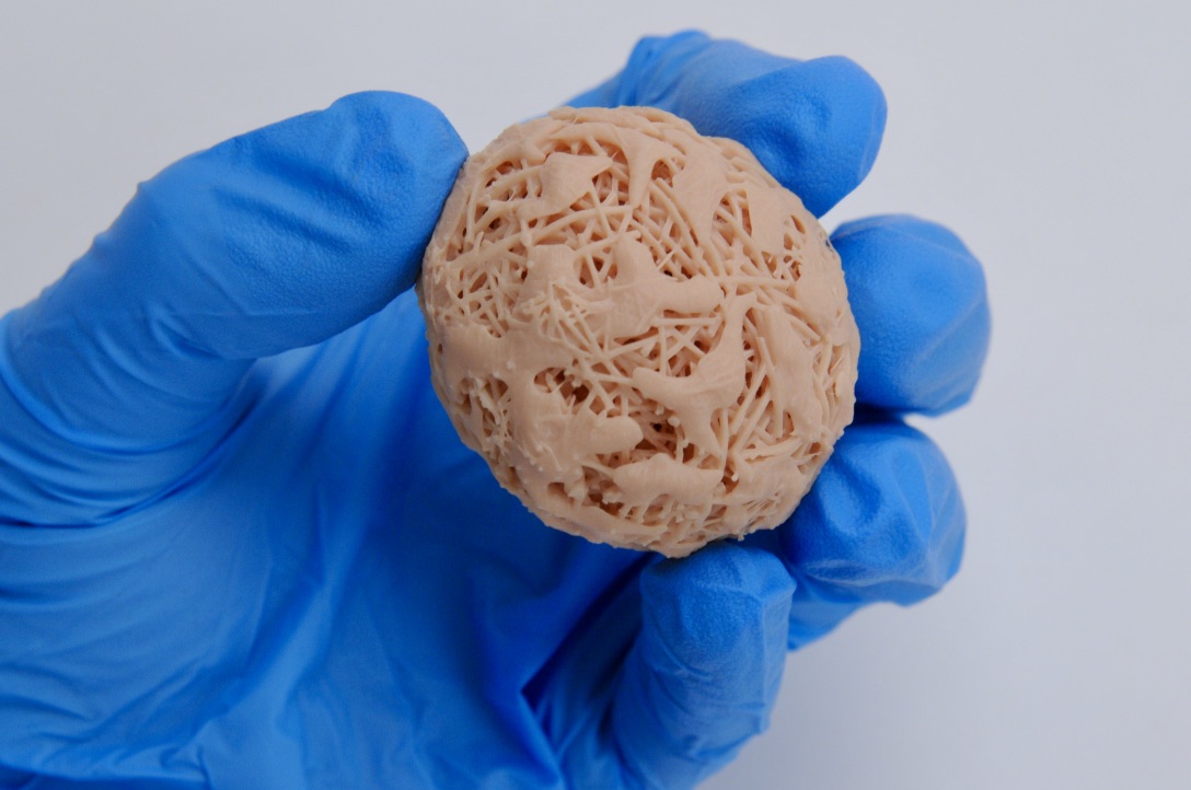
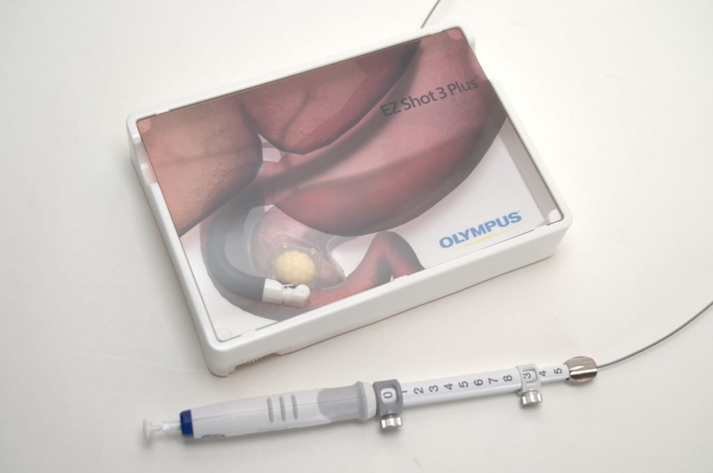
Instead, we created a compact, portable product composed of two pre-drilled suture deployment hole “rings.” By using a model design that was non-specific to an anatomical structure or joint, the physician was able to envision the device being utilized at any joint.
An anatomical demonstration makes sense for products used to target a specific body structure but would have limited the scope of this particular device. Additionally, the model was designed to allow for many (36 to be exact) practice sites, which resulted in the need for drastically fewer demo models.
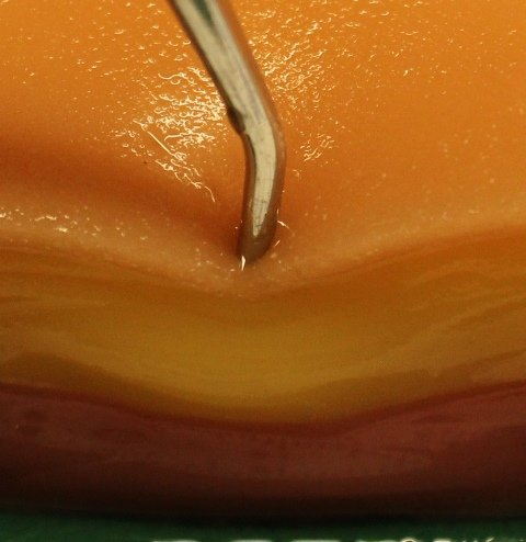
The outside holes in the bone block were open on the side that is up against the outer clear sleeve and provided excellent visibility of the mechanisms of the Suturefix Ultra using what we call the “ant farm” approach. The inside ring of holes was fully within the bone block and provided a secure anchor point for surgeons to conduct their own anchor pull test so they could feel the security they expected from the Suturefix anchor. This allowed for the best of both worlds in a convenient package.
Building a model that enhanced visualization, increased demonstration and practice applicability, and provided audio and tactile cues significantly augmented the ability to demonstrate the product’s effectiveness through the incorporation of multiple senses. Incorporating more sensory cues leads to improved recall and recognition of the product and the processes involved with using the product. This greatly benefits both the salesperson and the surgeon, which trickles down to ultimately improve patient outcomes.
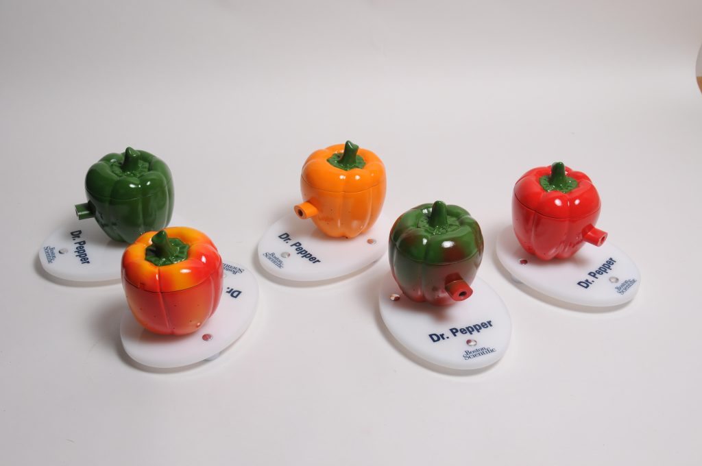
Dr. Pepper
For decades, medical device salespeople have used hollowed-out vegetables to represent internal body cavities. After all, vegetables provide a good deal of variation in size, contours, shapes, and textures. They’re also just…fun. Their vibrant colors and familiar, wholesome aromas are (literally) a breath of fresh air (especially compared to a cadaver).
They also allow for easy visualization and cleanup. However, as you can imagine, durability and reusability are not exactly their strong suits.
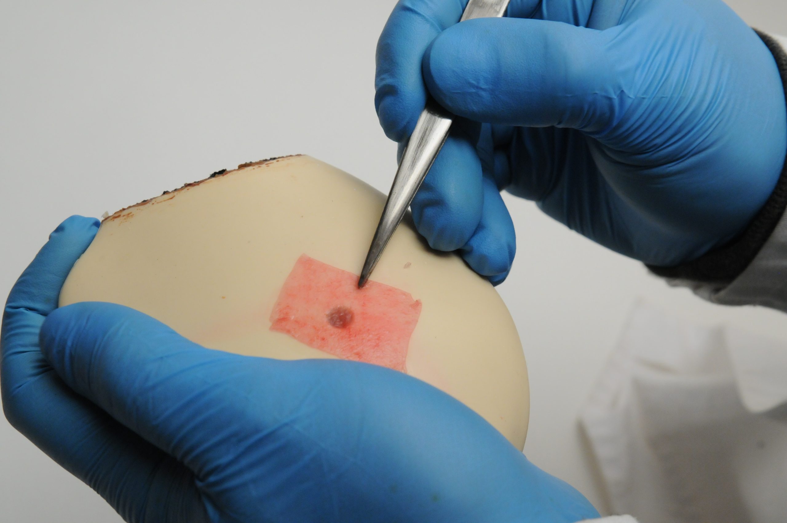
To intertwine the enjoyment and tradition of using a hollowed-out vegetable with the durability and beauty of a custom model, a forward-thinking client said, “Let’s use a pepper for the fun of it but make it into a model so it can be reused.” And with that came the birth of Dr. Pepper.
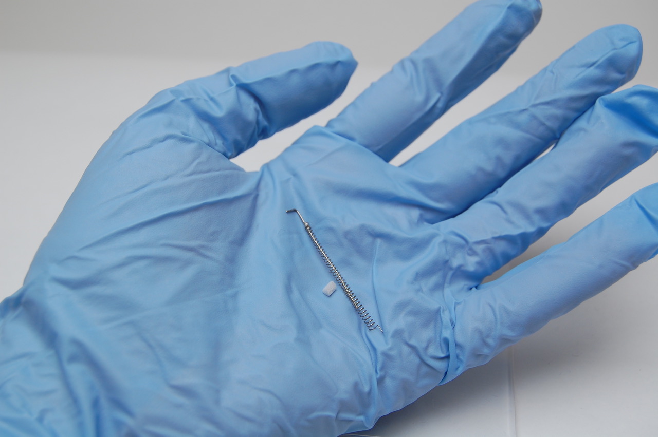
This Dr. Pepper is not a fizzy, delicious soft drink but rather the perfect blend of function and fun. Just as real peppers have been used in the surgical training space for decades, the purpose of this model is to allow surgeons to practice their wire manipulation skills.
Surgeons love the ability to conveniently hone their skills using a medium that is as playful as it is visually striking. Just as importantly, surgeons tend to be a spirited group of individuals who thrive on self-competition. The Dr. Pepper model addresses this by including a variety of colored dots with tiny entry points, each representing a different level of difficulty. As discussed in part 2 of this series, turning skills training into a game is not only amusing, but it also enhances skill retention and development compared to traditional modes of practice.
Salespeople benefit significantly from these factors because they can provide an experience that is professional and unmistakably memorable using a model that is durable and infinitely reusable. When the customer interaction shifts from a discussion to a hands-on experience, the product sells itself.
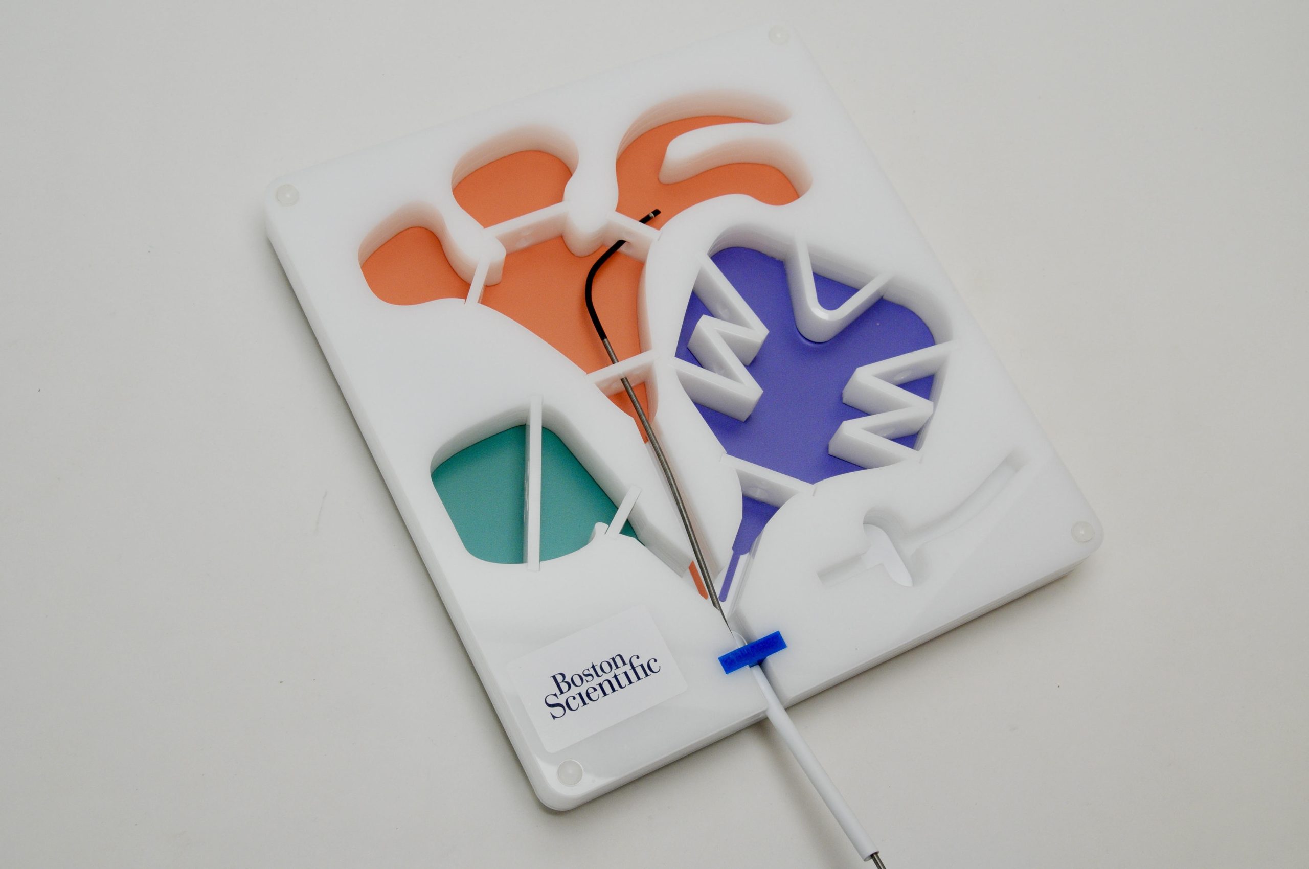
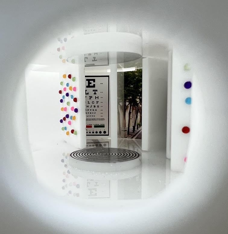
Just as the Dr. Pepper model turns skills training into a game while simultaneously providing a non-anatomical platform for enhanced visualization and surgical skill acquisition, so does The Funhouse.
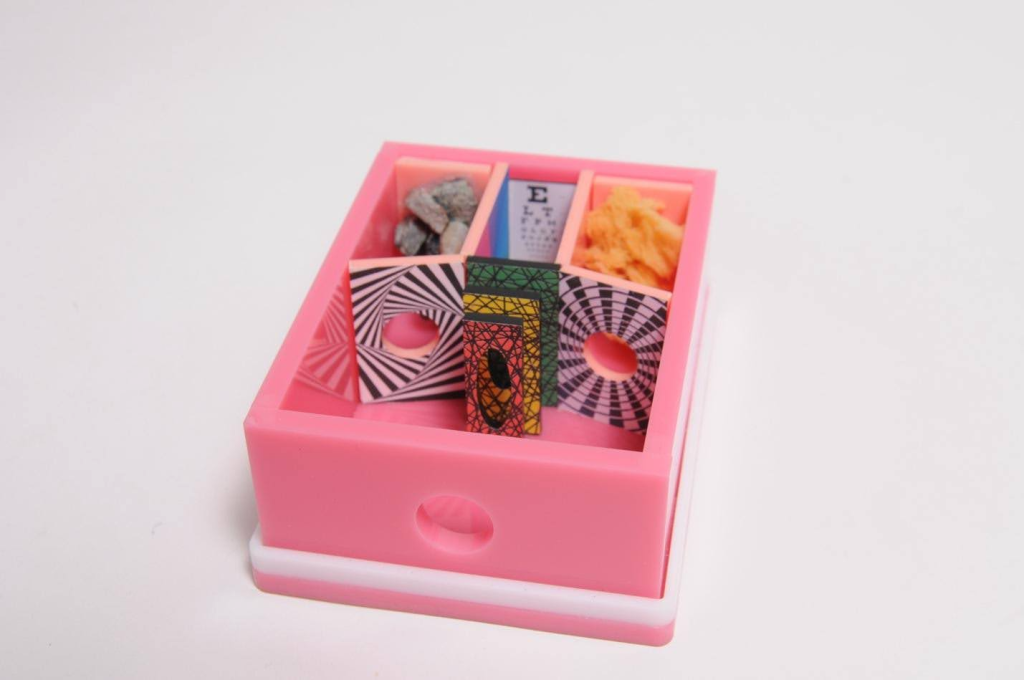
The Funhouse model was created as a vehicle to show scope illumination, image clarity, and color. But puttering around a basic demonstration using familiar anatomical landmarks doesn’t create as memorable of an experience as something novel and interesting. The consequence of predictability is the risk of getting lost in the noise of the competition or, worse, being bored. Our result is anything but boring.
The Funhouse includes rooms to highlight image contrast, depth of field, clarity of subtle texture using sponge and rock, and the ubiquitous eye chart. What is the smallest lettering you can see?
The model is small, not only for portability but to depict the volume of the body part the scope is intended to explore. This is important, as the engineers designed the light output to specifically match the organ’s volume so that if the model were too small or too large, it would not show off the scope’s performance.
As miraculous as The Funhouse looks from an artistic design perspective, it accomplished its functional goal. It allows clinicians to both experience the clarity of the company’s scope while also sharpening their navigation skills. These skills encompass the same immense precision and awareness needed when they have real patient lives in their hands.
The Funhouse
Wrap Up
Starting back in August of 2023, we set out to answer a simple question: Do all medical models need to represent anatomy? The answer to that question, in its most condensed form, is no. Medical demonstration models do not always need to represent anatomy to be functional, beautiful, and effective.
In fact, as the numerous examples of this four-part series have made abundantly clear, there are many times when non-anatomical models provide a better experience for both the customer and the salespeople.
The takeaway from all of this is what matters in the end is that an experience is provided. The canvas that experience is painted on won’t always be the same. This variability is the foundation of painting a picture that’s exciting, interesting, unique, and, most importantly, unforgettable.
So, thank you for perusing our medical demonstration model perspectives. We hope each of the four articles provided a different viewpoint and things to consider. And most of all, we hope you gained some valuable insights that you can apply now and in the future.